.png)

.png)
Objective: Transnasal endoscopic surgical treatments of the maxillary sinus (MS) and nasolacrimal canal (NLC) pathologies are very common. A detailed understanding of the anatomy of this region preoperatively affects the chances of surgical success. Advances in paranasal sinus computed tomography (PNSCT) have yielded a better understanding of the surgical anatomy.
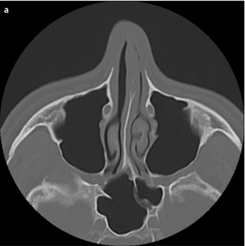
Material and Methods: The anatomical relationships between the medial and anterior walls of MS and NLC involving 81 patients (41 males and 41 females) were analyzed by means of PNSCT sections. The anatomical relationship of NLC with the MS anterior wall was evaluated and classified into unified and separate types. The separate-type NLC was further classified as protruding or nonprotruding according to the localization of the medial wall of the MS. In addition, the vertical height of the lower meatus and long and short diameters of the NLC were measured.
Results: The unified type was detected in 54 (32.9%) patients out of the total MSs of 164 patients. The distance between the anterior walls of the MS and NLC was 3.50 mm in females and 3.95 mm in males; this did not significantly differ between the genders. Further, 26 MSs were of the protruding type. The vertical height of the lower meatus was 10.75 mm in females and 12.15 mm in males. The long diameter of the NLC was 6.30 mm in women and 6.80 mm in men. The short diameter of the NLC was 4.85 mm in women and 5.00 mm in men.
Conclusion: The relationship between the NLC and MS shows anatomical variations. Understanding these variations before surgery can increase the success rate of endoscopic surgical procedures directed toward the medial wall of MSs.
Cite this article as: Akyüz S, Yıldırım FN, Ersöz N, Başak HS, Karaman CZ. Evaluation of Maxillary Sinus and Nasolacrimal Canal Relationship via Paranasal Sinus Tomography Imaging. Eur J Rhinol Allergy 2019; 2(3): 71-4.