.png)
.png)
Objective: The aim of this study was to compare the clinical radiological and pathological results of pathologies detected in isolated sphenoid sinus lesions.
Material and Methods: Six patients with sphenoid sinus lesions admitted to the Otorhinolaryngology and Head and Neck Surgery Clinic between July 2017 and October 2018 were retrospectively analyzed. Demographic characteristics, clinical findings, imaging findings, surgical treatment methods, and histopathological diagnosis were analyzed. The study was accepted by the Ethics Committee (Code, 2019/414; dated 02/09/2019).
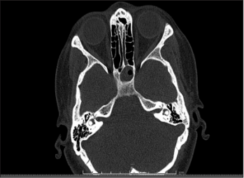
Results: The age range of the patients was between 18 and 71 years, the median age of the patients was 30.5 (between 18 and 71) years. The most common presenting complaints were headache (6/6), nasal obstruction (1/6), and postnasal discharge (1/6). Nasal endoscopic examination revealed purulent discharge in the middle meatus in two cases (2/6). One patient had septal deviation and concha hypertrophy (1/6). Examination was normal in two cases (2/6). Surgical intervention was performed in five patients (5/6). Follow-up was recommended in one patient (1/6) due to response to medical treatment. The pathological results of the patients who underwent surgery were sphenoid sinusitis in two cases, sphenoid sinus cyst in two cases, high-grade B cell lymphoma in one case, and chordoma in one case.
Conclusion: Isolated sphenoid lesions are the most common headache complaints, and the lack of findings on physical examination leads to delay in diagnosis. In cases of clinical suspicion and in cases resistant to medical therapy, imaging methods should be performed with computed tomography and/or magnetic resonance imaging. Surgery is the primary treatment approach, and definitive diagnosis is made by histopathological analysis.
Cite this article as: Aydın E, Akıdil AÖ, Ateş Evren G, Sayın İ, Güneş S, Yazıcı ZM. Isolated Sphenoid Sinus Lesions. Eur J Rhinol Allergy 2019; 2(2): 41-4.